


Table of Contents
- What Does a Gynecomastia Ultrasound Show?
- Do You Need an Ultrasound for Gynecomastia?
- Ultrasound or Mammogram: Which Test Comes First?
- What Happens During a Gynecomastia Ultrasound?
- Can an Ultrasound Tell Gynecomastia From Chest Fat?
- What Ultrasound Findings Mean You Need More Testing?
- How Does an Ultrasound Affect Gynecomastia Surgery Planning?
- What to Do Next
A gynecomastia ultrasound is a painless imaging test that shows whether the tissue behind your nipple is firm gland, soft fat, or something that needs a closer look. Most men do not need one, because a good physical exam answers the question. I am Dr. Babak Moeinolmolki, and I order an ultrasound when the exam is unclear or when something about a lump does not fit the usual pattern.
What Does a Gynecomastia Ultrasound Show?
A gynecomastia ultrasound shows whether breast gland tissue is present behind the nipple and what that tissue looks like. Early, active gynecomastia appears as a disc-shaped dark area under the areola. Older tissue looks brighter as scar-like fibrosis develops. Fat appears as a different, softer pattern.
A published review of imaging in gynecomastia describes florid, early-onset gynecomastia on ultrasound as a disc-shaped, hypoechoic area underlying the areola. The same review notes that the lesion becomes more echogenic as fibrosis develops, which matches what I feel on exam: recent tissue is often tender and rubbery, while long-standing tissue is firmer (PubMed 34033252).
The radiologist also looks at the edges of the tissue, the blood flow, and whether the pattern is symmetric. Findings are usually reported with a BI-RADS category, the standard scale that rates how suspicious an imaging finding is.
Do You Need an Ultrasound for Gynecomastia?
Most men with gynecomastia do not need an ultrasound. A history and a hands-on exam can usually confirm gland tissue. Imaging becomes useful when the exam is inconclusive, when a lump is hard, one-sided, or irregular, or when your surgeon wants to rule out another cause before surgery.
The imaging review cited above states that most male breast lesions can be diagnosed on clinical grounds and that imaging helps when the physical exam is inconclusive. Its stated goals are to confirm the diagnosis, separate true gynecomastia from pseudogynecomastia, exclude breast cancer, and look for a cause (PubMed 34033252).
My own rule is simple. If the chest is symmetric, the tissue is a soft disc under each nipple, and the story fits, I do not order imaging just to have a picture. If a man points to a hard lump on one side, has nipple changes, or has bloody discharge, the ultrasound comes first. You can read more about how I sort these cases in how gynecomastia is diagnosed and gynecomastia lump versus cancer.
Ultrasound or Mammogram: Which Test Comes First?
Ultrasound is generally the first imaging test for male breast enlargement. A mammogram can be added to confirm the finding or to look more closely at a suspicious area. Both tests can identify gynecomastia, and neither replaces a physical exam.
A surgical review of gynecomastia recommends ultrasound as the first-line imaging investigation, with mammography added to confirm the diagnosis (PubMed 11841077). The imaging review reports that both mammography and ultrasound are sensitive and specific for diagnosing gynecomastia and separating it from breast cancer.
| Feature | Ultrasound | Mammogram |
|---|---|---|
| Typical role | First imaging test | Added to confirm or clarify |
| Radiation | None | Low-dose X-ray |
| Gynecomastia appearance | Disc-shaped dark area under the areola | Subareolar density, often nodular, dendritic, or diffuse |
| Comfort | Gel and a handheld probe | Chest is compressed briefly |
| Time | About 15 to 30 minutes, varies by facility | About 15 to 30 minutes, varies by facility |
The three mammographic patterns named in the review are nodular, dendritic, and diffuse. They tend to correspond to early florid gynecomastia, long-standing fibrous gynecomastia, and gynecomastia linked to outside estrogen exposure.
What Happens During a Gynecomastia Ultrasound?
A technologist applies warm gel to your chest and moves a handheld probe over each side while you lie on your back. The test takes roughly 15 to 30 minutes, uses no radiation, and does not require sedation or fasting. Most men feel only light pressure.
You can usually eat, drink, and take your regular medications beforehand. Wear a two-piece outfit so only your shirt comes off. A radiologist reads the images and sends a written report. If you are a surgical candidate, I review the report alongside my exam, since the two together tell me far more than either one alone.
Tell the technologist about any tenderness, nipple discharge, or the exact spot where you feel a lump. That helps them scan the right area.
Can an Ultrasound Tell Gynecomastia From Chest Fat?
Yes, in most cases. Ultrasound can show whether there is a gland disc behind the nipple or only fatty tissue. This separates true gynecomastia from pseudogynecomastia, which matters because fat can shrink with weight loss while glandular tissue usually does not.
Distinguishing the two is one of the main goals of imaging in the review cited above. A second review of male breast imaging also lists pseudogynecomastia as an important mimic whose distinction affects the treatment plan (PubMed 32072386).
In my practice I still start with the hands-on check. The gynecomastia pinch test gives you a rough idea at home, and the article on pseudogynecomastia versus chest fat explains why the answer changes the treatment. An ultrasound settles the cases where the exam leaves doubt, such as a heavier chest where gland is hard to feel through the fat.
What Ultrasound Findings Mean You Need More Testing?
A solid, irregular, or one-sided mass, skin changes, enlarged lymph nodes, or an abnormal BI-RADS category usually lead to further testing. That can mean a mammogram, a needle biopsy, or referral to a breast specialist. Male breast cancer is uncommon, but it is the reason imaging exists.
The imaging review advises that when clinical findings suggest malignancy or imaging is inconclusive, histological confirmation should be sought. A needle biopsy samples the tissue so a pathologist can examine it.
The symptoms that raise my concern most are a hard fixed lump, a lump off to one side of the nipple, bloody nipple discharge, nipple retraction, or skin dimpling. If you have any of these, do not wait for a surgical consultation. See the guidance in nipple discharge in men and get examined promptly.
How Does an Ultrasound Affect Gynecomastia Surgery Planning?
An ultrasound helps confirm that the problem is gland tissue and rules out other causes of a lump before surgery. It does not choose the operation. The surgical plan depends on the physical exam, chest fat, skin quality, and your goals, and the technique varies by case.
The imaging review notes that imaging contributes to therapeutic decision-making. In practice, a clear report that shows gland tissue supports an operation that removes the gland and shapes the chest, usually with liposuction when fat is present. Tissue removed during surgery can also be sent to pathology, and I discuss that at the consultation.
Underlying causes matter too. The review reports that an underlying condition is found in roughly 45 to 50 percent of adult men with gynecomastia, including medications, systemic disease, obesity, and hormone disorders. That is why I look at the causes of gynecomastia before recommending anything. When surgery is the right step, the gynecomastia surgery guide walks through what to expect.
What to Do Next
If you have chest enlargement and are unsure whether it is gland or fat, start with a physical exam. Bring your medication list and a note about when the change began. Ask whether imaging is needed in your case instead of assuming it is. If a lump is hard, one-sided, or paired with discharge, get it checked soon.
To discuss your own chest, you can request a consultation through our male breast reduction page. Outcomes and recovery vary by case, and I will tell you plainly whether surgery makes sense for you.
Dr. Babak Moeinolmolki, MD, FACS, is double board-certified through the American Board of Cosmetic Surgery (ABCS) and the American Board of General Surgery. Any surgeon you consider should have credentials you can verify with their certifying board. This article is educational and does not replace an exam by your physician.
* Illustrative images on this page are models, not actual patients. Real patient results are shown in our Before & After gallery.
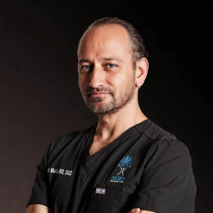
Dr.Babak Moeinolmolki
LA Cosmetic Surgeon Dr. Moein is board-certified by the American Board of General Surgery.